
By: Ross W. Nash, DDS
Provided by: Ed Matthews – Sure Business Logic
Introduction:
Single tooth edentulism is a pathology frequently treated with dental implants. However there are many cases where implants are not the ideal treatment plan in which case a minimally invasive bridge may be better indicated for the patient. First Fit technology offers an ideal solution for these cases. The technology was developed by Dr. Cyrus Tahmasebi and it is an innovative system that uses 3D printed guides to precisely prepare teeth and to deliver a crown or bridge or multiple veneers in one visit.
Procedure overview:
A First Fit bridge is a fixed dental restoration with a minimally invasive preparation using 3D printed guides based on a digital design.
Full arch upper and lower PVS impressions or a scan are sent to the First Fit lab. First Fit software creates a preparation design as well as a final restoration design for the clinician to approve. Then 3 D printed guides are made as well as the final restoration which is milled.
Case presentation:
A patient presented missing a mandibular first premolar. She knew that the other teeth were moving but could not afford an implant. She also did not want to have to wear a removable device. To give her a minimally evasive option, I proposed a FirstFit zirconia bridge which would require only small inlay preparations on the abutment teeth. These would be prepared at the time of seating.
Full upper and lower impressions were taken with a polyvinyl impression material. A bite registration was also taken. These were sent to the lab along with pre-operative photographs and an x-ray showing both abutment teeth. The lab fabricated the restoration and the preparation guide. These were returned to my office. The pre-operative image is shown in Figure 1 and the preparation guide can be seen in Figure 2. The zirconia bridge can be seen photographed on a mirror surface in Figure 3 and on the printed model in Figure 4.
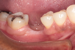
Figure 1
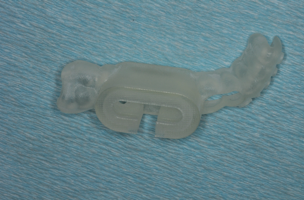
Figure 2
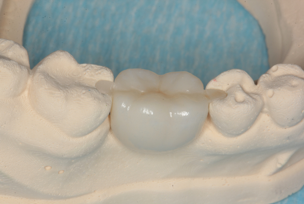
Figure 3
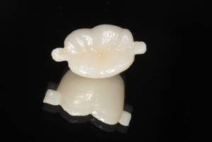
Figure 4
At the delivery appointment, the guide was seated and the special handpiece was inserted to prepare the two inlay rests with the diamond bur provided by the lab (Figure 5). The prepared rest seats can be seen in Figure 6. The bridge was bonded to place and can be seen in Figure 7. A radiograph of the FirstFit bridge in place is shown in Figure 8.
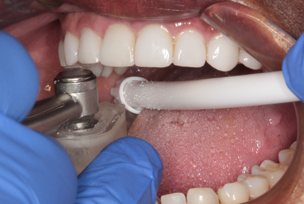
Figure 5
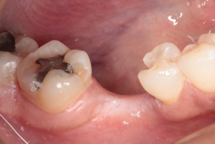
Figure 6
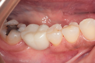
Figure 7
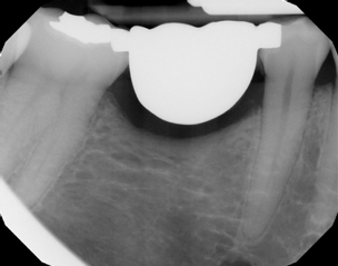
Figure 8
Conclusion:
By using a First Fit procedure, we were able to replace a patient’s mandibular first molar in a minimally invasive way allowing for other procedures in the future should they be needed or desired.
All dental photography by Ross W. Nash, DDS
To Learn more about First Fit for (DSO) Dental Service Organizations, visit: https://firstfit.com/. To Learn more about the Nash Institute, visit: http://www.thenashinstitute.com.
To schedule a lecture or seminar with one of our clinicians, specialized for (DSO) Dental Service Organizations, email – [email protected] or visit: https://www.surebusinesslogic.com/


 at the 2019 GNYDM")


